Follow us
Latest News & Events


Digital Pathology in ICCA Research: How KFBIO KF-PRO-400 Enables Biomarker Discovery
Introduction: Improving ICCA Research Through Digital Pathology
Intrahepatic cholangiocarcinoma (ICCA) is a rare and aggressive type of liver cancer.
Patients with ICCA often face limited treatment options and poor long-term outcomes. Therefore, finding reliable biomarkers is essential for personalized treatment and better patient management.
In recent years, immunotherapy has provided new opportunities for cancer treatment. However, patient responses can vary significantly.
As a result, researchers need more accurate methods to understand tumor characteristics and predict treatment outcomes.
Digital pathology is becoming an important tool in this process. By combining high-resolution imaging, image analysis and quantitative assessment, digital pathology helps researchers explore complex tumor features more efficiently.
A recent study published in Liver Cancer (DOI: 10.1159/000547326) investigated immune and histopathological biomarkers in ICCA. The study explored how the tumor microenvironment affects prognosis and response to neoadjuvant immunotherapy.
To support this research, the team used the KFBIO KF-PRO-400 Digital Slide Scanner.
The scanner provided high-resolution digitization of histopathological slides. Therefore, researchers could perform more consistent and detailed analysis of tissue features.
Study Overview: Connecting Immune Profiles with Clinical Outcomes
The research team analyzed two groups of ICCA patients.
The first group included patients who underwent surgical resection without previous treatment.
The second group included biopsy samples collected before neoadjuvant therapy (NAT).
Researchers used multiplex immunofluorescence and histopathological scoring to evaluate important tumor features.
These features included:
- CD8+ T-cell density
- Regulatory T-cell (Treg) density
- Tumor-infiltrating lymphocytes (TILs)
- Tumor budding (TB)
Together, these indicators helped researchers understand the relationship between immune activity, tumor structure and patient outcomes.
Key Findings: Immune Biomarkers Predict Patient Outcomes
The study identified several important findings:
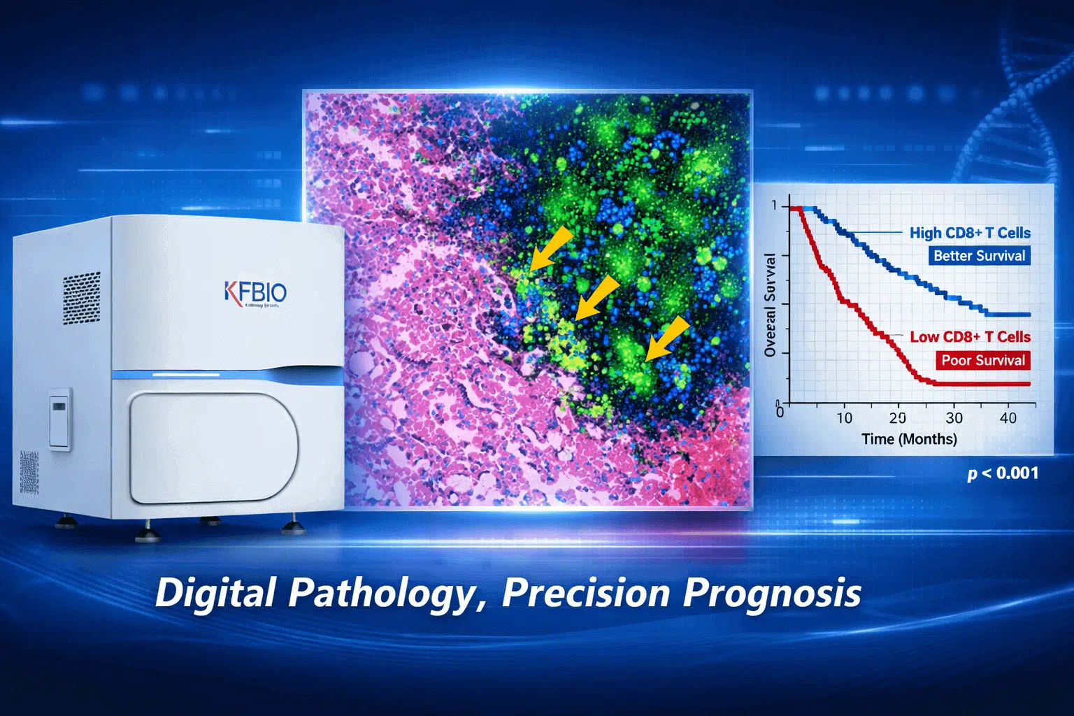
Higher CD8+ T-Cell Infiltration Improved Survival
Patients with higher levels of CD8+ T cells showed better overall survival outcomes.
This finding suggests that stronger immune activity may contribute to better treatment responses.
Higher Treg Density Was Linked to Poorer Outcomes
In contrast, increased regulatory T-cell density was associated with worse clinical outcomes.
Therefore, evaluating different immune cell populations can provide valuable information for patient risk assessment.
TILs and Tumor Budding Help Predict Immunotherapy Response
The study also found that TIL density and tumor budding were strong independent predictors of treatment response.
These results highlight the importance of analyzing both immune features and tumor architecture.
More importantly, they demonstrate the value of quantitative pathology analysis in modern cancer research.
The Role of Digital Pathology in Modern Cancer Research
Traditional pathology relies heavily on manual slide examination.
Although experienced pathologists provide valuable expertise, manual evaluation can be time-consuming and may vary between observers.
Digital pathology provides a new approach.
By converting glass slides into high-resolution digital images, researchers can perform standardized analysis and share data more efficiently.
In this ICCA study, researchers scanned H&E-stained slides from biopsy and resection samples using the KFBIO KF-PRO-400 Digital Slide Scanner.
This process supported:
- Standardized slide evaluation
- Remote collaboration
- Quantitative analysis of tissue feature
- More reliable biomarker assessment
Furthermore, digital pathology allows researchers to analyze the relationship between tissue morphology and immune markers.
As a result, it supports the development of predictive models for personalized cancer treatment.
KF-PRO-400: Enabling High-Quality Digital Pathology Research
To perform reliable biomarker analysis, researchers need accurate and consistent digital images.
Therefore, high-quality whole slide imaging (WSI) plays an essential role in modern pathology research.
The KFBIO KF-PRO-400 Digital Slide Scanner provides a powerful imaging solution for pathology laboratories and research institutions.
The scanner enables researchers to digitize tissue samples with high resolution and excellent image quality.
It supports multiple imaging applications, including:
- Brightfield imaging
- Fluorescence imaging
- FISH imaging
With fast scanning performance and automated operation, the KF-PRO-400 helps laboratories improve research efficiency.
Moreover, its user-friendly design makes it suitable for both clinical environments and research applications.
Key Features of the KF-PRO-400 Digital Slide Scanner
The KF-PRO-400 is designed to support large-scale digital pathology workflows.
Its key features include:
High-Resolution Whole Slide Imaging
The scanner uses a 40x objective lens with a high numerical aperture (0.95).
This capability allows researchers to capture detailed tissue structures and cellular information.
As a result, it supports accurate analysis of complex pathological features.
Automated Slide Handling and Focus
The KF-PRO-400 provides automated slide loading and intelligent focusing.
Therefore, laboratories can reduce manual operation and improve workflow consistency.
This automation is especially valuable for research projects involving large numbers of tissue samples.
Flexible Software Compatibility
The scanner supports integration with major digital pathology image management systems.
This flexibility allows laboratories to connect scanning workflows with existing digital pathology platforms.
Furthermore, researchers can easily manage, review and share digital slides.
Compact Design for Laboratory Integration
The KF-PRO-400 combines advanced imaging performance with a compact design.
Therefore, it can be integrated into different laboratory environments without requiring major workflow changes.
Supporting Biomarker Discovery and Cancer Research
In the ICCA study, the KF-PRO-400 played an important role in tissue digitization.
The scanner converted histopathological samples into high-quality digital images for further immune and pathological analysis.
This process provided a reliable foundation for:
- Immune marker evaluation
- Tumor architecture analysis
- Quantitative pathology scoring
- Predictive model development
As digital pathology continues to advance, reliable imaging technology becomes increasingly important.
Without standardized and high-quality digital images, researchers cannot fully utilize advanced image analysis or AI-based tools.
Therefore, digital slide scanners like the KF-PRO-400 are becoming essential tools in biomarker discovery and translational cancer research.
Clinical Implications and Future Directions
The findings from this ICCA study demonstrate the growing value of digital pathology in precision oncology.
By analyzing biomarkers such as TILs and tumor budding, researchers can better understand patient responses and treatment outcomes.
In the future, digital pathology may help clinicians:
- Classify patient risk more accurately
- Select more suitable treatment strategies
- Develop personalized therapeutic approaches
Furthermore, digital pathology can work together with other technologies.
For example, combining genomic information with digital image analysis may provide deeper insights into tumor biology.
As a result, researchers can develop more comprehensive predictive models.
The Future of Digital Pathology in Precision Medicine
Digital pathology is transforming cancer research and clinical workflows.
Traditional microscopy provides valuable diagnostic information. However, digital pathology adds new possibilities through data analysis, collaboration and artificial intelligence.
With advanced scanners such as the KFBIO KF-PRO-400, laboratories can build reliable digital pathology workflows.
These workflows support:
- Research collaboration
- Quantitative tissue analysis
- AI development
- Precision medicine applications
Looking ahead, digital pathology will continue to play an important role in oncology.
It will help researchers discover new biomarkers, improve treatment strategies and support better patient outcomes.
Conclusion: Advancing Cancer Research with Digital Pathology
This ICCA study demonstrates how digital pathology can improve cancer research.
By enabling detailed and quantitative analysis of tumor-immune interactions, digital pathology provides valuable insights into disease progression and treatment response.
The KFBIO KF-PRO-400 Digital Slide Scanner provided the imaging foundation for this research.
Through high-resolution scanning, automated operation and flexible integration, the scanner supports reliable pathology analysis and biomarker discovery.
As digital pathology continues to evolve, its role in clinical research and personalized medicine will become even more important.
Ultimately, technologies such as the KF-PRO-400 will help laboratories move toward smarter, more efficient and data-driven pathology workflows.
References & Links:
-
Original study: Liver Cancer, DOI: 10.1159/000547326
-
Learn more about the KF-PRO-400 Scanner: https://www.kfbio.cn/kf-pro-400
-
Digital Pathology in Cancer Research: https://www.ncbi.nlm.nih.gov/pmc/articles/PMC7879852/